Watermelon Bacterial Rind Necrosis in Oklahoma
This year’s prolonged hot summer and droughts are already wreaking havoc on plants. We are now beginning to see outbreaks of some crop diseases that are not usually part of the disease landscape of Oklahoma. A watermelon disease that is known to occur in prolonged periods of drought is watermelon bacterial rind necrosis (WBRN, BRN or WRN, I prefer WBRN or BRN because of the ‘bacteria’; plus, recovered isolates associated with the disease have been shown to cause symptoms on other hosts, so I will use BRN). Interestingly, one of the earliest reports of an outbreak of BRN in the United States occurred after a period of drought in Georgia in 1925 (Gilbert and Artschwager, 1925). The disease is thought to be sporadic throughout the US, with Florida and Georgia reporting major outbreaks in 2011 and 2012. The disease has also been reported in California, Delaware, Hawaii amongst other states. BRN typically appears as a hard and dry browning of watermelon rind that rarely extends to the flesh. External symptoms are also rare, but misshapen fruits in melons with advanced stages of internal necrosis have been reported (Hopkins and Elmstrom, 1974). BRN is poorly understood, and strains of bacteria belonging to multiple species have been frequently isolated from infected lesions and shown to induce necrotic symptoms in laboratory inoculations.
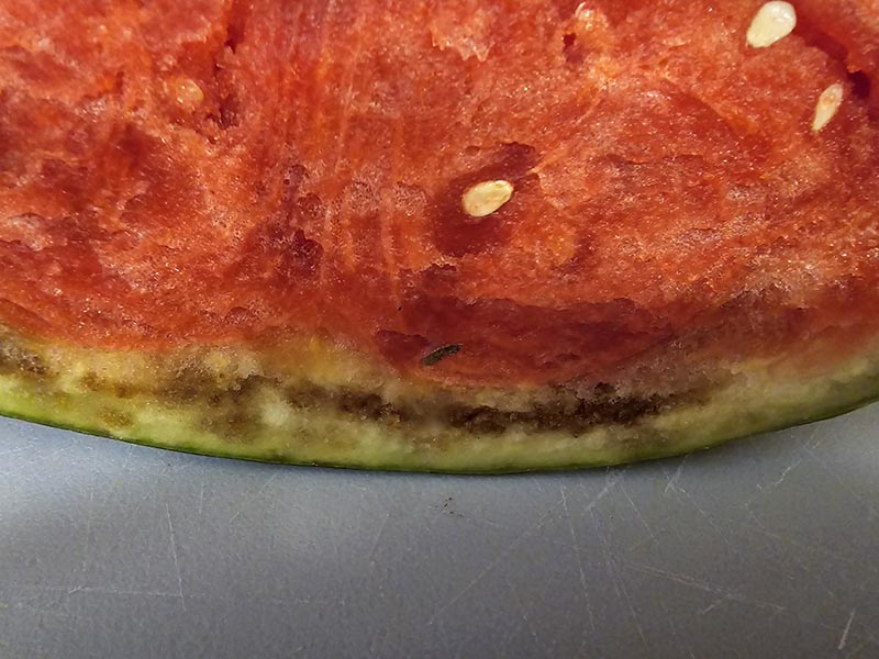
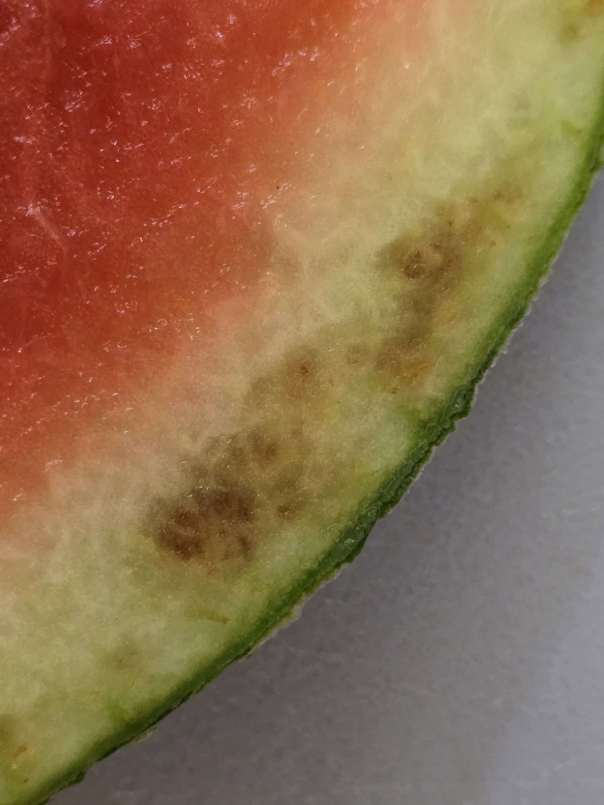
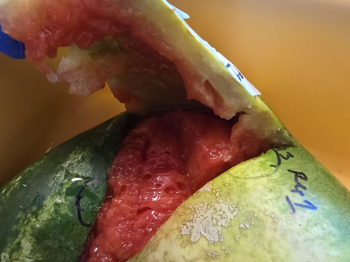
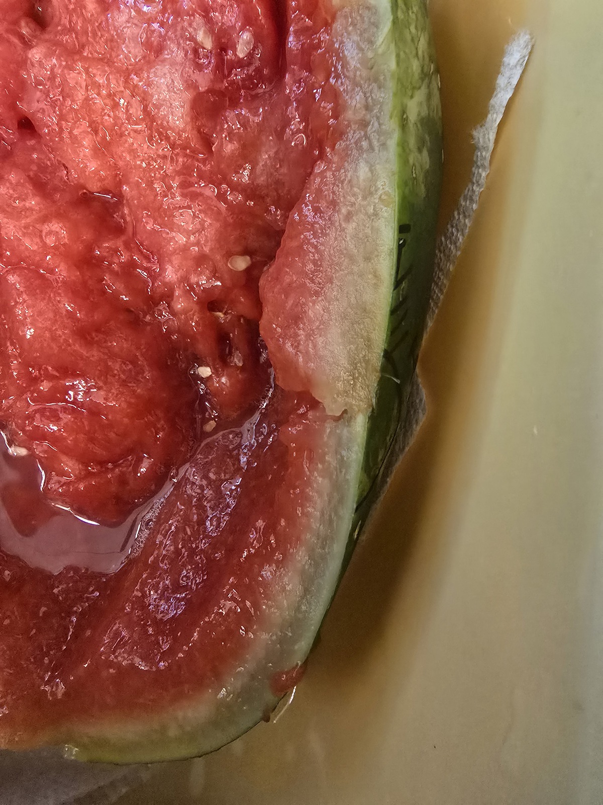
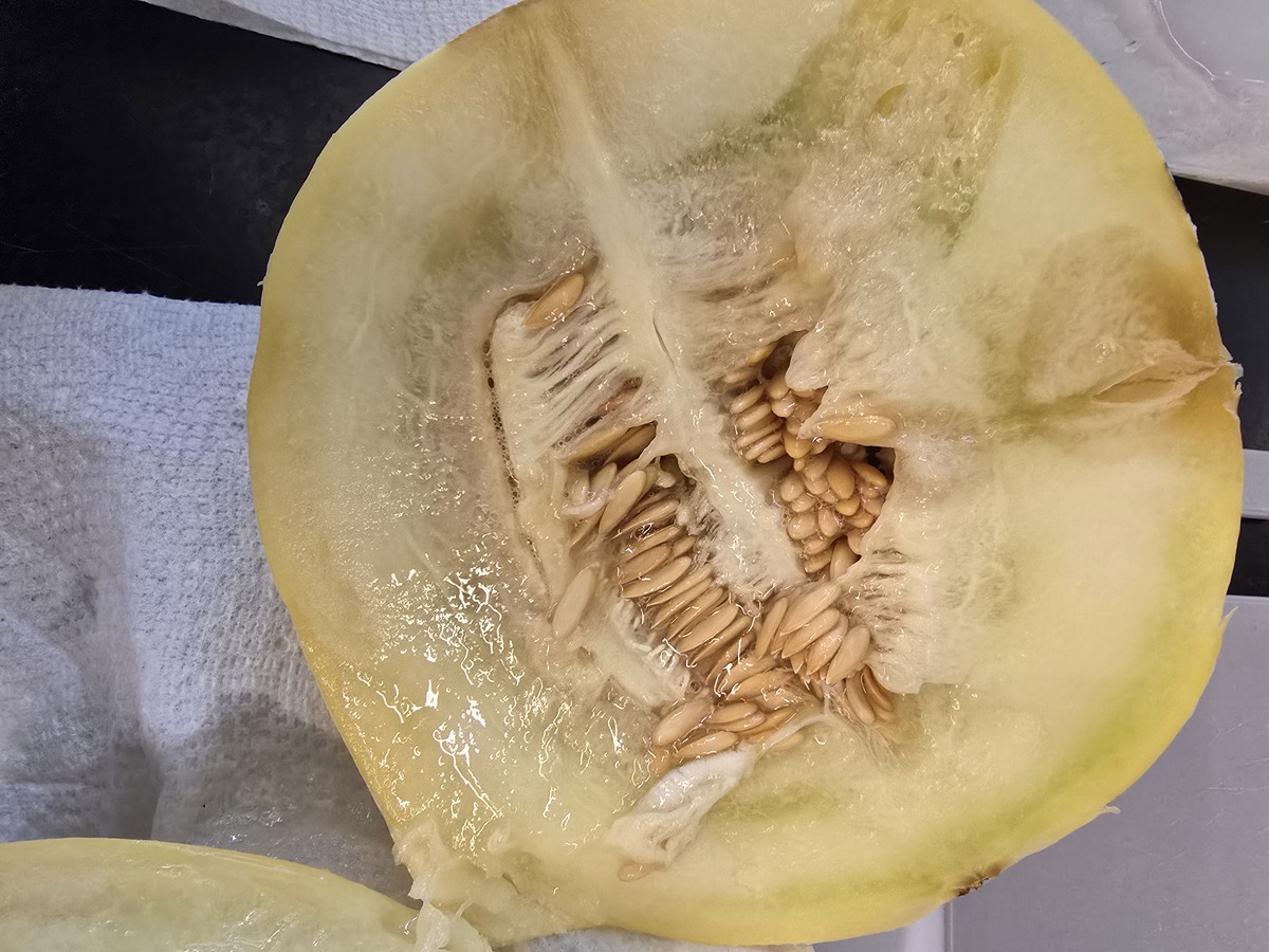
A watermelon grower within the state recently observed corky dark brown symptoms on the rind of the seedless watermelon variety, Fascination. Rind necrosis symptoms were observed on the seedless variety but not the seeded pollinator. We observed similar symptoms when we cut open watermelon samples submitted to us by the grower (Figures 1a and b). Subsequently, we isolated pure cultures of at least five different bacterial species from submitted samples. When grown on King’s Medium B and visualized under ultraviolet light, some of the recovered strains showed fluorescence typical of a pseudomonad (Figure 2). Sequencing of the 16S ribosomal RNA gene identified strains belonging to Pantoea, Erwinia, Pseudomonas, Bacillus and Acinetobacter species. Strains of all five bacterial groups caused similar symptoms when inoculated on the mini seedless watermelon variety, PureHeart and melon variety, honeydew. Water-soaked lesions rapidly led to a collapse of the internal tissues, including the rind within four days (Figure 3a, 3b and 3c). Although there are no described symptoms associated with BRN on the leaves, we recovered a similar set of bacteria from submitted watermelon leaves. Various studies have isolated different sets of bacteria from BRN symptoms, and it is thought to be a disease complex. Interestingly, Squash Vein Yellowing Virus (SqVYV) causes similar symptoms on watermelon. We did not test the samples for SqVYV.
Watermelons infected with BRN necrosis are not marketable. It is not confirmed if bacteria recovered from BRN symptoms have any human health consequences. However, Enterobacter cloacae, Klebsiella pneumoniae and Kosakonia cowanii were identified in BRN and rot lesions on melon in Turkey (Ozdemir, 2021). These species are part of the normal human gut flora and have only been reported to cause diseases in immunocompromised individuals. As Oklahomans learn to live with prolonged hot summers and droughts, this may become an important disease in the state. Unfortunately, not so much work has been done on epidemiology and management of the disease and this should be a focus. If you observe similar symptoms in your watermelon field, please contact your local extension office.
Figures 1a-b. Symptoms of watermelon bacterial rind necrosis on watermelon samples submitted by a grower.
Figure 1a.
Figure 1b.
Figure 2. Some of the recovered isolates on King’s Medium B fluorescing under Ultraviolet light.
Figures 3a-c. Symptoms on watermelon (cultivar “Pure Heart”) and melon (cultivar “honeydew” ) inoculated with recovered bacterial species (at 108 cfu/ml) that were isolated from submitted watermelon samples. Pictures were taken 4 days after bacterial inoculation, but symptoms may have developed earlier over the weekend.
Figure 3a.
Figure 3b.
Figure 3c.
References
Gilbert, W. W., and Artschwager, E. (1925). Watermelon internal browning. Phytopathology 15:119-121.
Hopkins, D. L., and Elmstrom, G. W. (1977). Etiology of watermelon rind necrosis. Phytopathology 67: 961-964.
Ozdemir, Z. (2021). Identification of Enterobacteriaceae members and fluorescent pseudomonads associated with bacterial rind necrosis and rot of melon in Turkey. Eur J Plant Pathol (2021) 160:797–812. https://doi.org/10.1007/s10658-021-02282-z

